
Summary
- Sesamoid injuries are extremely common, especially in high-arched individuals.
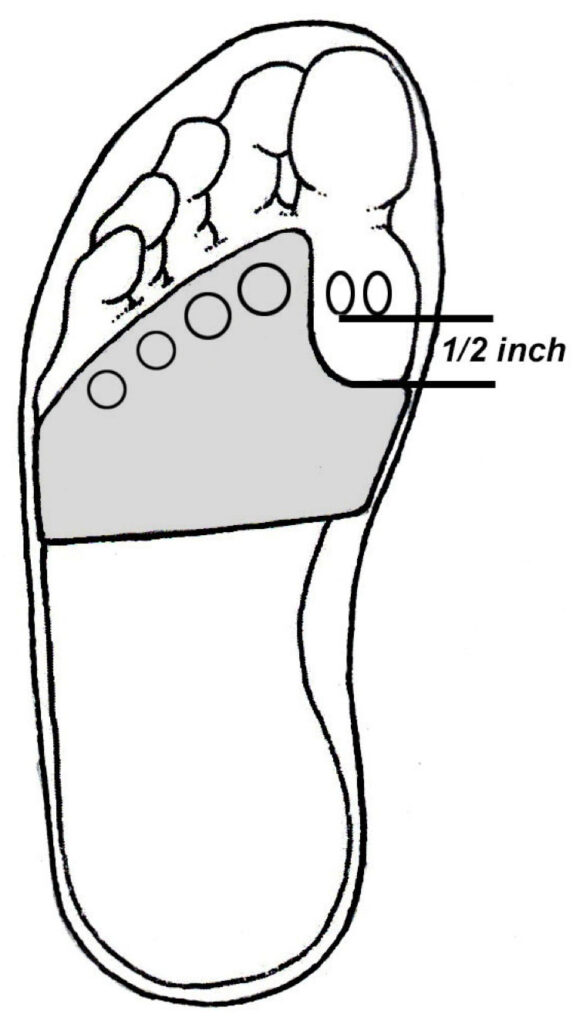
- The new Peel and Stick Sesamoid Balance is thicker than most sesamoid balances, and is made of a blend of urethane rubber, PPT, and synthetic suede designed to significantly reduce pressure beneath the first metatarsal head.
The word sesamoid is Latin for “sesame seed,” and these small bones are located inside specific tendons where they improve mechanical efficiency by pulling the tendon farther away from the joint’s axis of motion. The classic example of a sesamoid is the patella, which improves mechanical efficiency of the quadriceps by more than 50% (1). The foot also possesses important sesamoids. Located beneath the first metatarsal head, the tibial and fibular sesamoids (also known as the medial and lateral sesamoids) are situated in the tendon of the flexor hallucis brevis muscle where they are separated by a small groove on the bottom surface of the metatarsal head (Fig. 1). The sesamoids are invaluable during the gait cycle as they increase force production beneath the great toe during propulsion, which improves stability and transfers pressure away from the smaller central metatarsals. Ferris et al. (2) prove that forceful downward movement of the great toe during push-off reduces central metatarsal pressure by as much as 30%, and may even play an important role in lessening the potential for metatarsal stress fractures and interdigital neuromas.
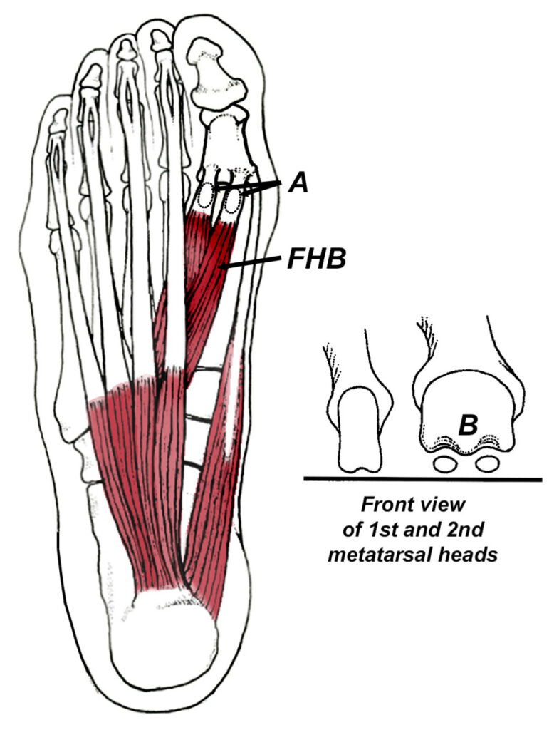
The sesamoids are separated by a bony crest on the bottom of the first metatarsal head (B).
Unfortunately, because of their location, the sesamoid bones are subjected to tremendous compressive forces. This is especially true in people with high arches. Force plate analysis shows that people with high arches have a greater distribution of pressure centered beneath their first and fifth metatarsal heads, while people with neutral and low arches have greater pressure distribution beneath the central metatarsals (Fig. 2).
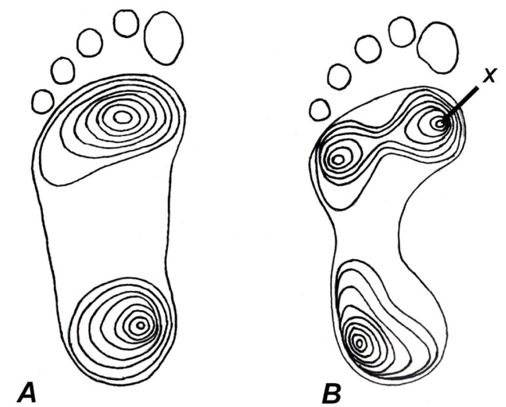
Notice how pressure is centered directly beneath the medial sesamoid in individuals with high arches (X).
To protect people with high arches from being injured, one of the most important things you can do is to shift pressure away from the sesamoid bones. The new Peel and Stick Sesamoid Balance was designed to do just that (Fig. 3). Made from a blend of urethane rubber, PPT foam, and synthetic suede, the Peel and Stick Sesamoid Balance attaches directly to the bottom of an insole in just a few seconds.
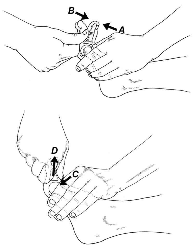
In addition to the Peel and Stick Sesamoid Balance, people with chronic sesamoid pain should be evaluated for contracture in the flexor hallicus brevis muscle. This important muscle tightens in response to sesamoid pain, and the resultant contracture pulls the sesamoids up into the base of the first metatarsal head as the big toe bends during propulsion. Contracture in flexor hallucis brevis is evaluated by measuring the degree of dorsiflexion in the first MTP (Fig. 4).
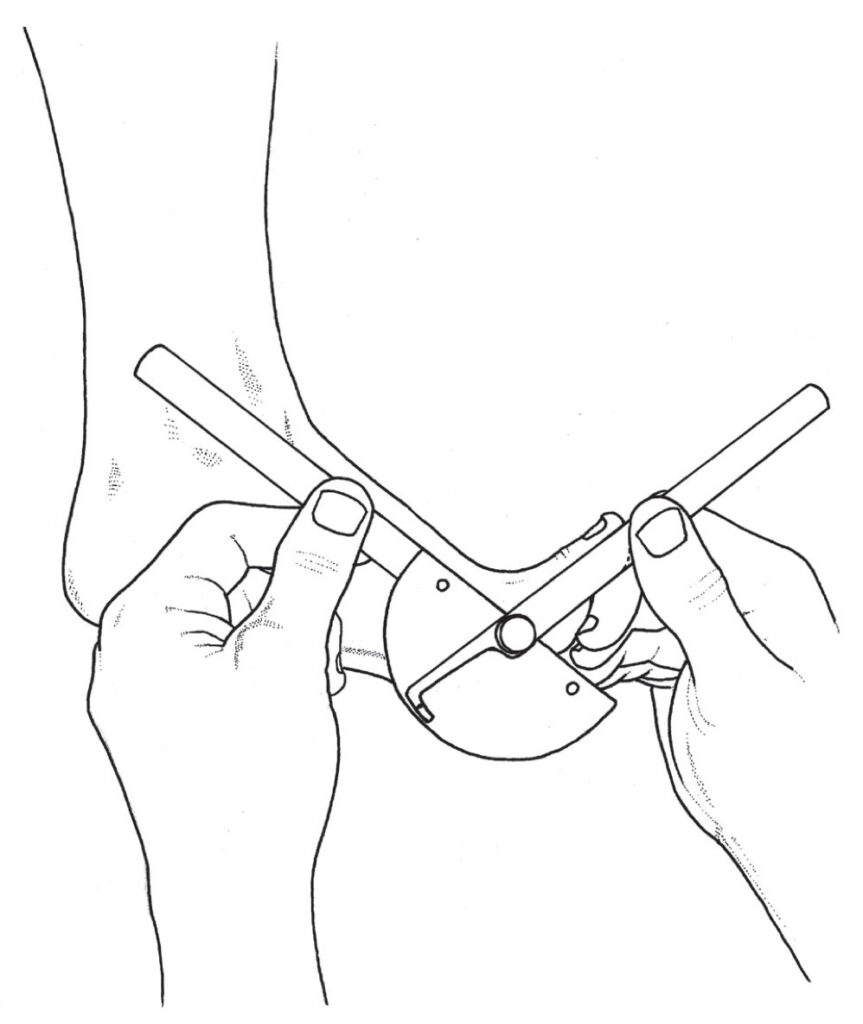
When upward motion of the big toe is limited (compare to the range available on the uninjured side), the mobilization illustrated in figure 5 effectively restores motion in the first MTP. Of course, home stretches for maintaining motion in the first MTP should be emphasized. Between manual therapy and the Peel and Stick Sesamoid Balance, even the worst sesamoid injuries can typically be resolved without the need for surgical referral.
References:
- Fulkerson J. Disorders of the patellofemoral joint. 3rd edition. Baltimore (MD): Williams & Wilkins;1997.
- Ferris L, Sharkey N, Smith T, et al. Influence of extrinsic plantar flexors on forefoot loading during heel rise.
Foot Ankle. 1995;16:464-473.