
In any given year, nearly one in 25 athletes will tear their anterior cruciate ligament (1). More commonly, one in 10 recreational athletes develop patellofemoral pain annually, and once diagnosed, more than 90% will continue to suffer with chronic knee pain years later (2). To make matters worse, individuals with ACL tears and/or patellofemoral pain are significantly more likely to develop arthritis later in life (3).
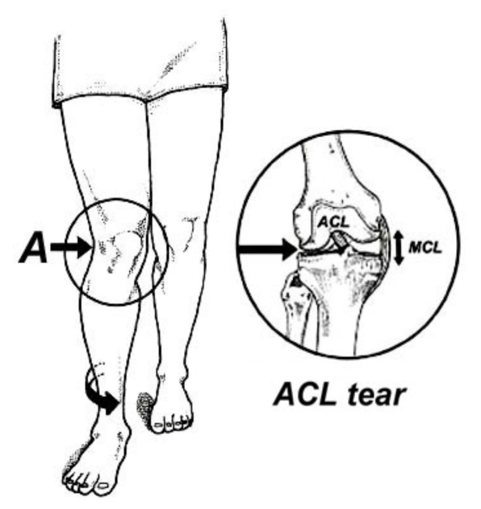
Fortunately, new research shows it is possible to identify at risk athletes with a simple muscle test using a handheld dynamometer (4). Because anterior cruciate ligaments are associated with excessive hip internal rotation and valgus collapse of the knee during stance phase (Fig. 1), researchers from Iran theorized that athletes prone to tearing their anterior cruciate ligaments would to present with weak hip abductors and rotators, and would therefore be less likely to muscularly control their lower extremities while participating in sport (4).
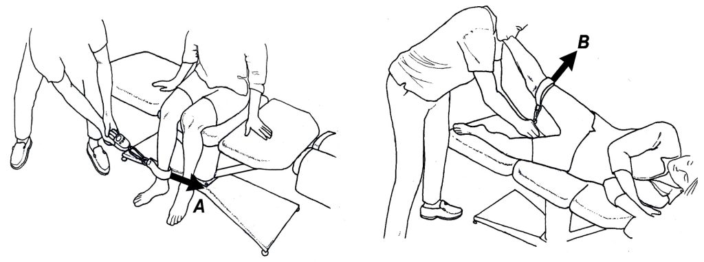
To test their theory, these researchers followed 531 competitive athletes (138 females and 363 males) participating in various sports for one season after measuring hip strength with a handheld dynamometer. The specific muscle tests are illustrated in figure 2. Athletes were classified as “strong” if they could generate more than 20% and 35% of their body weight in their hip rotators and abductors, respectively. Conversely, athletes were classified as “weak” if they fail to meet these minimum hip strength requirements.
Using the strength measurements based on body weight, these researchers demonstrated that at the end of the season, fewer than 1% of the strong athletes suffered ACL injury, while 7% of athletes with weak hips tore their ACLs. Having a weak piriformis muscle was especially correlated with increased rates of ACL tears. This is consistent with prior research (5) confirming that isolated piriformis weakness strongly correlates with the development of a wide range of lower extremity injuries, including patellofemoral pain syndrome in runners (Fig. 3), and ankle injuries in basketball players.

Knowing in advance which athletes are prone to ACL and/or retropatellar injuries is incredibly important because there are proven exercise interventions that markedly reduce injury rates. For example, Gilchrist et al. (6) was able to reduce ACL injuries by 70% in 1,435 Division I college female athletes by having them perform a series of simple agility drills. More recently, Baldon et al. (7) had women with chronic retropatellar pain perform specific functional exercises to strengthen their hips and knees. Not only did these exercises reduce pain and improve function, they also reduced valgus collapse of the knee while exercising. Interestingly, these authors compared the functional exercises with conventional physical therapy exercises targeting the hips and knees (such as leg presses, seated knee extensions, and straight leg raises), and only the functional movement patterns improved three-dimensional movement patterns.
Considering the long-term physical and emotional consequences associated with ACL and/or patellofemoral injuries, every athlete should be tested preseason to quantify his or her hip strength ratios. Weak athletes should be encouraged to perform specific training protocols to reduce their potential for injury. Measuring hip rotator/ abductor strength with a handheld dynamometer is easy, reliable, and subsequent testing during re-examination can help evaluate response to a specific exercise intervention. Additional information, including a video of various hip exercises, is available on www.humanlocomotion.org.
References:
- Moses B, Orchard J, Orchard J. Systematic review: annual incidence of ACL injury and surgery in various populations. Res Sports Med. 2012;20(3-4):157-179.
- Stathopulu E, Baildam E. Anterior knee pain: a long-term follow-up. Rheumatology (Oxford). 2003;42(2):380-382.
- Utting MR, Davies G, Newman JH. Is anterior knee pain a predisposing factor to patellofemoral osteoarthritis? Knee. 2005;12(5):362- 365.
- Khayambashi K, Ghoddosi N, Straub R, et al. Hip Muscle Strength Predicts Noncontact Anterior Cruciate Ligament Injury in Male and Female Athletes: A Prospective Study. Am J Sports Med. 2015;(44):355-361.
- Leetun D, Ireland M, Willson J, et al. Core stability measures as risk factors for lower extremity injury in athletes. Med Sci Sports Exerc. 2004;36:926–934.
- Gilchrist J, Mandelbaum H, et al. A randomized controlled trial to prevent noncontact anterior cruciate ligament injury in female collegiate soccer players. Am J Sports Med. 2008;36:1476-1483.
- Baldon R, Serrao F, Silva R, Piva S. Effects of functional stabilization training on pain, function, and lower extremity biomechanics in women with patellofemoral pain: a randomized clinical trial. J Orthop Sports Phys Ther. 2014;44(4):240-251.